Common Types of Medical and Diagnostic Imaging
Medical imaging is a vast field of study and an important tool in modern medicine. It allows your doctor to look inside your body, identify what’s causing your symptoms and determine the best course of action to treat you. Medical imaging services can be broken down into three general categories: X-rays, laboratory tests and scans. Each type of diagnostic imaging has its own specific benefits and limitations, but they can all play an essential role when diagnosing a medical condition or determining the best treatment option for you. The key to understanding diagnostic imaging is knowing the different types of it available and the common uses for each type. This article explores some of the most common types of diagnostic imaging used in modern medicine.
X-rays
X-rays are still the most common type of medical imaging services used today, despite the fact that they have been replaced by other types of imaging as they became more widely available. They are widely used to diagnose orthopedic injuries and tumors, as well as certain cancers, such as cervical and prostate. They are also frequently used for the following: – Examining the structure and function of the bones and soft tissues, such as muscles and tendons and ligaments. This can help your doctor determine the underlying cause of your pain. – Identifying broken bones, kidney stones and gallstones, urinary stones, and other abnormalities inside your body. – Diagnosing how well a metal implant, such as a knee or hip replacement implant, is working. – Evaluating joint problems, such as osteoarthritis, in the knee. – Evaluating the function of your heart and lungs.
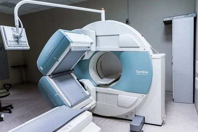
Image Source: Pixabay
Ultrasound
Ultrasound uses sound waves to create images of areas inside your body. It is used to assess conditions inside your body, including the following: – Differentiating between normal and abnormal tissue. For example, you might have an ultrasound to check if you have a kidney stone or to see if your gallbladder is inflamed, as it could be causing your stomach pain. – Examining your blood vessels, to see if they are causing inflammation or if you have an infection. – Examining your fetus for signs of problems, such as whether the fetus may be too big for the uterine cavity. – Monitoring your pregnancy. You may have ultrasound scans to check the growth of your fetus. – Evaluating joint problems or injuries, such as ruptured disks in the back, to see if you need surgery. – Evaluating the function of your heart and lungs.
MRI (magnetic resonance imaging)
An MRI scan uses powerful magnets and radio waves to create detailed images of your body. It is used to evaluate injuries and conditions that may cause pain, such as the following: – Examining the structure of your brain, liver and other organs to determine if there is damage. – Studying your ligaments, muscles, tendons and joints to see if they are damaged or scarred and to determine if surgery is required. – Examining your heart, lungs and other major organs to rule out problems. – Studying your blood vessels to determine if they are blocked or damaged, which could be a sign of a certain medical condition.
CT Scan
A CT scan uses x-rays to create cross-sectional images of your body. CT scans are used to diagnose a variety of conditions and injuries, including the following: – Evaluating blood flow in the body, such as in the brain, heart, or lungs. – Determining the size, location and type of tumors or other abnormalities found in your body. – Studying your internal organs, such as the brain, heart, stomach and intestines, to rule out any problems. – Examining your bones, such as your spine, bones in your hand and wrist, shoulder, and ankle. CT scans can show how your bones are arranged and how they are related to each other and your organs.

Comments